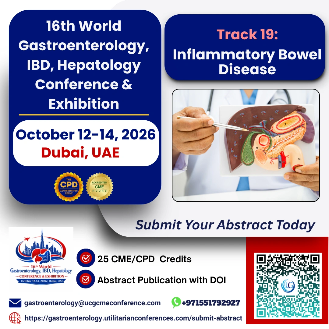


Gastroenterology is the study of the normal function and diseases of the...

What is Hepatology?
Hepatology is a specialized branch of medicine focused...

About of the Gastrointestinal Radiology
Gastrointestinal Radiology is a subspecialty within radiology that focuses on the imaging, diagnosis, and sometimes interventional treatment of diseases affecting the gastrointestinal (GI) tract, which includes organs like the esophagus, stomach, intestines, liver, pancreas, gallbladder, and bile ducts. This field is essential for detecting, monitoring, and guiding the treatment of a wide range of conditions, from common issues like gallstones and appendicitis to complex diseases such as cancers of the GI tract.
Key Components:
1. Advanced Imaging Techniques:
Utilizes sophisticated imaging modalities such as X-rays, CT scans, MRI, and ultrasound to obtain detailed images of the GI organs.
Specialized procedures like fluoroscopy and contrast studies (e.g., barium swallow and enema) help visualize the movement and structure of the GI tract in real-time.
2. Diagnostic Excellence:
GI radiologists are highly trained to interpret subtle findings in these images, which can be critical in diagnosing conditions early and accurately.
They play a vital role in cancer detection, identifying tumors and assessing the spread of disease.
3. Interventional Radiology:
Some gastrointestinal radiologists are also trained in minimally invasive procedures that treat conditions without the need for open surgery.
Techniques like ERCP (Endoscopic Retrograde Cholangiopancreatography) and MRCP (Magnetic Resonance Cholangiopancreatography) are used to diagnose and sometimes treat biliary and pancreatic duct conditions.
4. Collaborative Care:
GI radiologists work closely with gastroenterologists, surgeons, oncologists, and other specialists to ensure that patients receive comprehensive care.
Their insights often guide the clinical management of complex GI diseases, influencing treatment decisions and patient outcomes.
Gastrointestinal Radiology is an integral part of modern medicine, providing the imaging expertise necessary for the effective diagnosis and treatment of GI disorders.
Risks of GI radiology?
Gastrointestinal Radiology, while generally safe and highly effective in diagnosing and sometimes treating GI conditions, does come with certain risks. These risks vary depending on the specific imaging modality or procedure used. Here's an overview of potential risks associated with GI radiology:
1. Radiation Exposure:
X-rays and CT Scans: These imaging techniques involve exposure to ionizing radiation, which, in high doses or with repeated exposure, can increase the risk of cancer. However, modern equipment and protocols are designed to minimize this exposure as much as possible.
Cumulative Radiation: Patients who undergo multiple imaging studies over time, particularly CT scans, may accumulate higher levels of radiation exposure, potentially increasing their long-term cancer risk.
2. Contrast Agents:
Allergic Reactions: Some GI radiology procedures use contrast agents (e.g., barium, iodine-based contrast) to enhance image clarity. These can cause allergic reactions in some patients, ranging from mild (itching, rash) to severe (anaphylaxis).
Nephrotoxicity: Iodine-based contrast agents used in CT scans can be harmful to the kidneys, especially in patients with pre-existing kidney conditions, leading to contrast-induced nephropathy.
Aspiration Risk: During procedures like a barium swallow, there's a small risk that the contrast material could be aspirated (inhaled) into the lungs, particularly in patients with swallowing difficulties.
3. Procedure-Related Risks:
Endoscopic Retrograde Cholangiopancreatography (ERCP): This procedure, which combines endoscopy and fluoroscopy, carries risks such as pancreatitis, infections, bleeding, and perforation of the GI tract.
Perforation: Some imaging procedures, particularly those involving endoscopy or the use of contrast agents under pressure (like a barium enema), carry a small risk of perforating the GI tract.
Infection: Any procedure that involves insertion of instruments into the body, such as ERCP or biopsy under imaging guidance, carries a risk of infection.
4. Claustrophobia and Discomfort:
MRI and CT Scans: These procedures may cause discomfort or anxiety in patients who are claustrophobic, as they require staying still in an enclosed space for a period of time.
Bloating and Discomfort: Procedures involving ingestion of contrast materials or air insufflation (e.g., during a barium enema or CT colonography) can cause temporary bloating, discomfort, or cramping.
5. False Positives/Negatives:
Misinterpretation of Results: Although rare, there is a risk of false positives (finding an issue that is not actually present) or false negatives (missing a condition that is present), which can lead to unnecessary anxiety, additional testing, or delayed treatment.
6. Delayed Allergic Reactions:
Some patients may experience delayed allergic reactions to contrast agents, developing symptoms hours to days after the procedure.
7. Pregnancy Risks:
Fetal Exposure: Pregnant women are generally advised to avoid radiologic procedures involving ionizing radiation unless absolutely necessary, as exposure can pose risks to the developing fetus.
While these risks exist, the benefits of GI radiology often far outweigh them, especially when it comes to the early detection and treatment of serious conditions. Medical professionals take precautions to minimize these risks, such as using the lowest possible radiation dose, screening for allergies, and ensuring procedures are only performed when necessary.
When is GI radiology needed?
Gastrointestinal Radiology is needed in various clinical scenarios to diagnose, evaluate, and sometimes guide the treatment of conditions affecting the gastrointestinal (GI) tract. Here are common situations when GI radiology is essential:
1. Evaluation of Persistent Symptoms:
Abdominal Pain: Persistent or severe abdominal pain can indicate various conditions, such as appendicitis, pancreatitis, or bowel obstruction. Imaging helps determine the cause.
Chronic Diarrhea or Constipation: Imaging can help identify underlying structural or functional issues in the intestines, such as inflammatory bowel disease (IBD) or tumors.
Unexplained Weight Loss: Significant, unexplained weight loss may suggest malignancy or other serious conditions, prompting imaging studies to identify the cause.
Dysphagia (Difficulty Swallowing): Barium swallow or esophagography can be used to assess swallowing difficulties and detect abnormalities like strictures or tumors.
2. Cancer Detection and Staging:
Suspected GI Cancers: Imaging is crucial for diagnosing cancers of the esophagus, stomach, pancreas, liver, colon, and rectum. Techniques like CT, MRI, and PET scans are often used.
Staging of Known Cancers: Once a GI cancer is diagnosed, imaging helps determine the stage by assessing the tumor size, involvement of nearby structures, and presence of metastasis.
3. Inflammatory and Infectious Diseases:
Crohn's Disease and Ulcerative Colitis: MRI and CT enterography can provide detailed images of the intestines, helping to assess the extent and severity of these inflammatory conditions.
Pancreatitis: CT or MRI can help diagnose acute or chronic pancreatitis, identify complications, and guide treatment.
Hepatitis and Liver Cirrhosis: Ultrasound, CT, or MRI may be used to evaluate liver inflammation, fibrosis, and the presence of complications like portal hypertension or hepatocellular carcinoma.
4. Evaluation of Obstructive Conditions:
Bowel Obstruction: X-rays, CT scans, or fluoroscopic studies (like a barium enema) are used to diagnose and locate the site of obstruction in the intestines.
Biliary Obstruction: Ultrasound, MRCP, or ERCP can assess blockages in the bile ducts caused by gallstones, tumors, or strictures.
5. Preoperative and Postoperative Assessment:
Preoperative Planning: Imaging is often required before GI surgery to map out the anatomy and assess the extent of disease.
Postoperative Complications: After surgery, imaging may be needed to check for complications like leaks, infections, or bowel obstruction.
6. Trauma Assessment:
Abdominal Trauma: In cases of blunt or penetrating trauma to the abdomen, CT scans are often used to evaluate for injuries to the liver, spleen, intestines, and other organs.
7. Screening and Surveillance:
Colon Cancer Screening: Techniques like CT colonography (virtual colonoscopy) are used as non-invasive alternatives to traditional colonoscopy for colon cancer screening.
Surveillance of Known Conditions: Patients with a history of polyps, liver cirrhosis, or previous cancers may undergo regular imaging to monitor for recurrence or progression.
8. Evaluation of Jaundice:
Jaundice: Imaging such as ultrasound, CT, or MRCP is used to evaluate the cause of jaundice, which could be due to liver disease, gallstones, or bile duct obstruction.
9. GI Bleeding:
Active GI Bleeding: When a patient presents with symptoms of GI bleeding (e.g., blood in stool or vomit), imaging can help locate the source of the bleeding, especially when endoscopy is inconclusive.
10. Unexplained Anemia:
Chronic GI Blood Loss: Imaging studies may be needed to find the source of chronic blood loss leading to anemia, which can sometimes be due to small or hard-to-detect GI lesions.
Gastrointestinal Radiology is a critical tool for the early diagnosis and management of many GI conditions, ensuring timely and appropriate treatment.
Sub Track: Chronic GI Blood Loss, Active GI Bleeding, GI Bleeding, Evaluation of Jaundice, Jaundice, Screening and Surveillance, Trauma Assessment, Preoperative, Postoperative Assessment, Evaluation of Obstructive Conditions, Inflammatory, Infectious Diseases, Cancer Detection and Staging, Evaluation of Persistent Symptoms, Abdominal Pain, Chronic Diarrhea, Constipation, Unexplained Weight Loss, Suspected GI Cancers, Staging of Known Cancers, Pancreatitis, Hepatitis, Liver Cirrhosis, Bowel Obstruction, Biliary Obstruction, Delayed Allergic Reactions, Claustrophobia and Discomfort, Aspiration Risk, Nephrotoxicity, Allergic Reactions, Radiation Exposure, X-rays and CT Scans, Cumulative Radiation, Interventional Radiology, Diagnostic Excellence, Advanced Imaging Techniques.